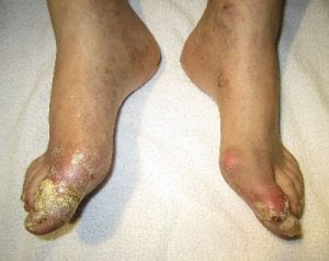
Gouty tophi are usually noticed under the skin, and are not usually painful until they burst through the skin, or become infected.
We tend to ignore them in the early stages, and concentrate more on relieving the pain from acute gout flares.
But is this wise?
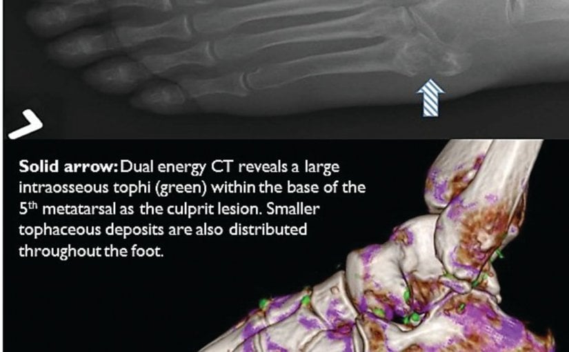
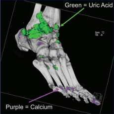
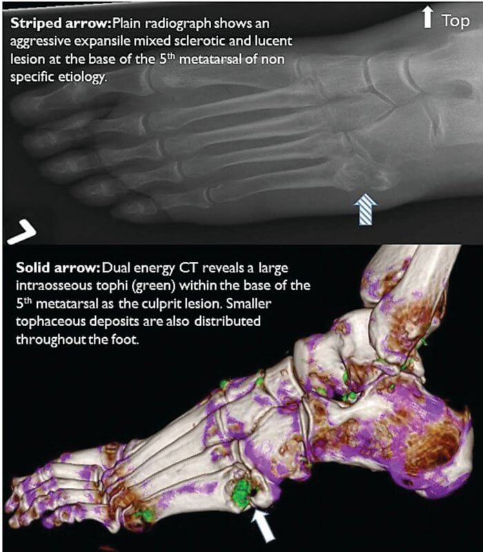
Now that technology allows us to see tophi growing into cartilage, tendons, and bones, we can see it is not wise. I’ve added some more important 2014 information to this 2010 article about early detection of joint damage from new gout scanning technology. First let us see how this technology shows early tophi leading to bone erosion and severe, painful, joint damage.
Gouty Tophi & Bone Erosion Revealed
It has been a few years since improvements in scanning techniques allowed us to view what is happening inside joints. Over 3 years ago, I quoted from Dalbeth [1]:
There is a strong relationship between bone erosion and the presence of intraosseous tophus. These results strongly implicate tophus infiltration into bone as the dominant mechanism for development of bone erosion and joint damage in gout.
Gouty Tophi & Bone Erosion Reviewed
A significant part of Dalbeth’s, and related research, is the observation that DECT reveals urate deposits much more effectively than any other technique, to the point that joints often show uric acid deposits prior to any physical or inflammatory indication of gout.
There is a commonly held belief that asymptomatic hyperuricemia (high uric acid with no gouty arthritis symptoms) does not need to be treated. This DECT research prompts me to believe that this policy needs to be reconsidered. As I said in my DECT review earlier this year [2]:
Lowering uric acid is more important than generally recognized. The policy of waiting for
several acute gout attacks before commencing urate lowering therapy is outdated, especially
given these clear indications that tendon damage takes place prior to acute gout flares.
Fortunately, this view is beginning to change amongst the more enlightened members of the rheumatology profession. Black and colleagues [3] note:
Utilization of imaging studies in order to accurately diagnose, monitor progression or treatment response, and assess clinical outcomes is increasing rapidly
However, they do add a cautionary note:
Limited initial availability will likely continue the role of DECT to those individuals with diagnostic uncertainty or access to larger imaging centers.
Gouty Tophi & Bone Erosion Restrained
Perhaps the cautionary note is contagious.
It is disappointing to see that whilst recent professional advice recognizes the value of DECT it fails to acknowledge its importance as a diagnostic technique. That advice also fails to recognize the implications on asymptomatic hyperuricemia.
Schumacher & Weaver [4] give with the one hand…
Recent studies show that DECT scans reveal significantly more areas of urate deposition than a physical examination. […] But if these newer imaging techniques can identify MSU crystal deposition prior to bone erosion or joint destruction, they offer promise as a noninvasive alternative for diagnosing gout in the earliest stages of the disease. In addition, they may be used in the future to monitor the progression of gout and the effectiveness of ULT.
… but take with the other:
The consensus standard of care today is that asymptomatic hyperuricemia should not be treated.
Perhaps there will come a day when DECT and other imaging techniques are common enough to dispel the myth that high uric acid is only dangerous when accompanied by acute gout flares. Until then, I recommend you do all you can to maintain your uric acid at a safe concentration below 6mg/dL (0.36 mmol/L).
Gouty Tophi & Bone Erosion: 2014 Update
The availability of gout scanning technology has improved now that Toshiba have released DECT for gout. Recent professional rheumatology guidelines now give us a framework for earlier effective gout treatment.
You can discuss how to avoid bone erosion from gouty tophi at those two gout forum links. If you prefer more facts about tophi, you will find them in my Tophi Guidelines.
Leave Gouty Tophi & Bone Erosion to browse the Tophi Guidelines.
Gouty Tophi & Bone Erosion Related Topics
Please remember: to find more related pages that are relevant to you, use the search box near the top of every page.
Common Terms: Ann. Rheum. Dis., gouty tophi definition, tendon, Tophi
Other posts that include these terms:
- Why are tophi important to gout sufferers?
- Gouty Tophi In Finger Bone
- Uric Acid, Devil’s Claw, and Gout
- Is Milk Good for Gout?
- Best Tophi Picture shows greatest Tophi Treatment
- Penis Tophi
- Gout & Rheumatoid Arthritis
- Doctor, do I have gout?
Gouty Tophi & Bone Erosion References
- Dalbeth, Nicola, Barnaby Clark, Kate Gregory, Greg Gamble, Timothy Sheehan, Anthony Doyle, and Fiona M. McQueen. “Mechanisms of bone erosion in gout: a quantitative analysis using plain radiography and computed tomography.” Annals of the rheumatic diseases 68, no. 8 (2009): 1290-1295.
- Taylor, Keith. “DECT For Gout Diagnosis.” GoutPal.com (June 2010). Keith Taylor DECT For Gout Diagnosis Archive.
- Black DF , Glazebrook K , Bongartz T , Matteson EL , Manek NJ , Leng S , Fletcher JG, McCollough C. “Dual-Energy Computed Tomography for the Evaluation of Gout and Calcium Crystal Deposits.” Conference Paper: Radiological Society of North America 2009 Scientific Assembly and Annual Meeting. Gouty Tophi Revealed With DECT Archive.
- Schumacher, H. and A. Weaver. “A Specialty Monograph Focusing on Core Principles in the Diagnosis and Management of Gout and Hyperuricemia.” (2010). Corpus ID (S2CID): 73330596. Gout And Hyperuricemia Archive.
Please give your feedback
Did this page help you? If yes, please consider a small donation. Your donations help keep GoutPal's gout support services free for everyone.
If not, please tell me how I can improve it to help you more.
- YouTube
- The gout forums.